
Glial tumors are a broad group of tumors arising from the glial cells of the brain. They are classified as low-grade or high-grade, and the aim of treatment is to remove as much tumor as possible without worsening the patient's condition.
What are glial tumors (astrocytomas)?
Glial tumors are a broad group of tumors arising from the glial cells (astrocytes) of the brain. They are the second most common tumors originating in the brain.
Although they can occur at almost any age and in any region, they are encountered more often between the ages of 30 and 40 and over 60.
They can be broadly divided into low-grade and high-grade tumors. Molecular and genetic research in recent years has shown that this tumor type has different genetic characteristics from patient to patient and requires individualised treatment.
High-grade tumors
These grow more quickly and can be life-threatening if not treated early. Because high-grade tumors are likely to recur, radiotherapy (radiation treatment) and chemotherapy (drug treatment) are required after surgery if the patient's general condition and the tumor's size and location allow.
Low-grade tumors
These may remain silent and unchanged in size for a long time or grow very slowly. Because most of them do eventually grow, may extend into critical areas of the brain, may reach a large size and may transform into a more malignant form, surgery is usually the first-line treatment. Less commonly, for small tumors in very high-risk locations, biopsy followed by radiotherapy and chemotherapy may be used.
Symptoms
They may present with headache, seizures (fainting or convulsions as seen in epilepsy), numbness or weakness on one side of the body, drowsiness, slowing, changes in behaviour and personality, and speech disturbance.
Diagnosis
Magnetic resonance imaging is essential for diagnosis. The tumor type and grade are confirmed by pathological and molecular examination of tissue obtained at surgery or biopsy.
Treatment Approach
The aim of surgery is maximal tumor removal without worsening the patient's clinical condition. When this is achieved, the recurrence rate may decrease and the time to recurrence may be prolonged.
Today, high-technology tools such as intraoperative MRI (MRI performed during surgery), neuronavigation, fluorescence-guided operating microscopes and ultrasonography allow these tumors to be removed more safely than in the past.
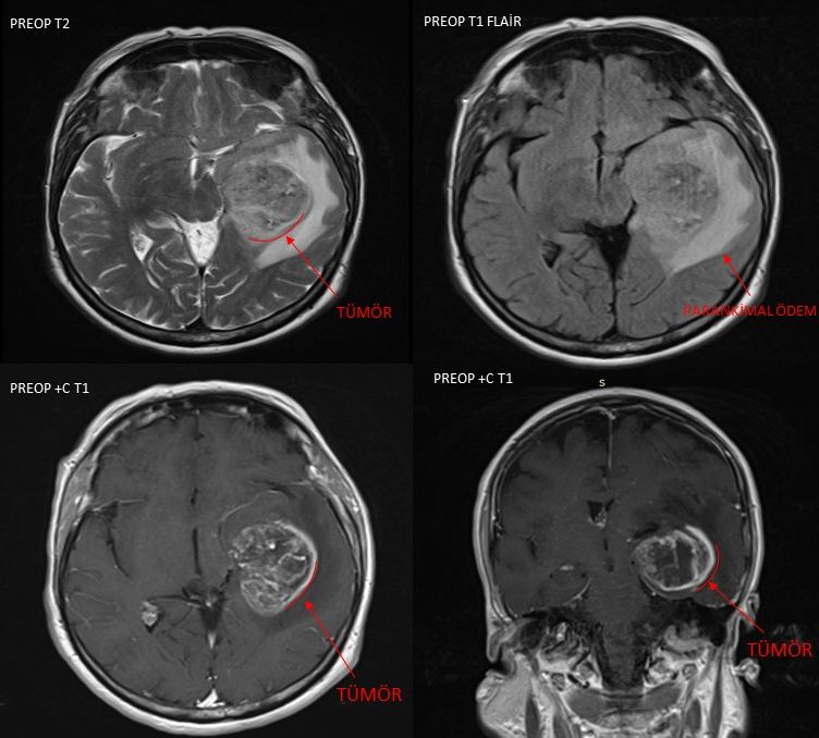
Preoperative MRI
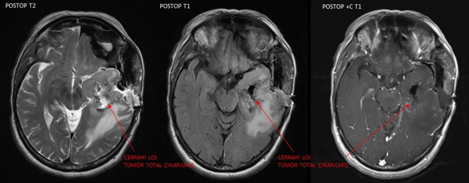
Intraoperative and postoperative MRI showing complete removal of the tumor.