
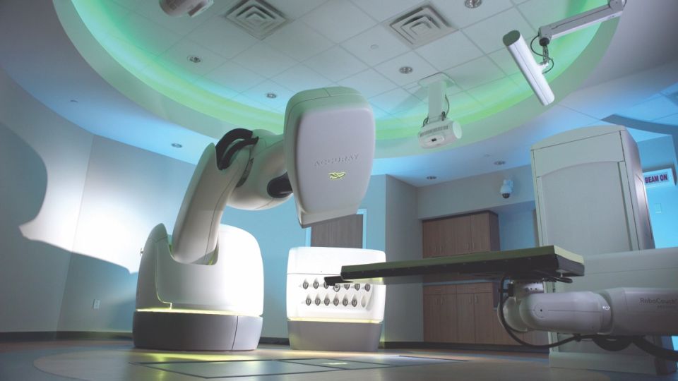
CyberKnife is a radiosurgery system that uses a linear accelerator mounted on a robotic arm to focus X-rays onto a target, and can be used for lesions both inside and outside the head.
CyberKnife is a radiosurgery system that delivers treatment by focusing X-rays generated by a linear accelerator onto a defined target.
Unlike Gamma Knife, it uses a mask rather than a frame, allows treatment over more than one session and can also be used outside the head. Because no frame is fixed to the skull, its precision (<1 mm) is not quite that of Gamma Knife, but it is of a level and quality that ensures safe treatment. CyberKnife combines an image-guidance system that locates and tracks the target throughout treatment with a computer-controlled robot, allowing tumors to be irradiated with high precision.
The robotic system coordinates a three-dimensional reconstruction of the skull, created from a previously acquired CT scan, with radiographs taken during treatment. A radiation source redesigned specifically for radiosurgery is integrated into the system: a lightweight 6 MV linear accelerator that moves along six coordinate axes on the robot. FDA clearance in 2001 opened the way for its use throughout the body.
Because it can deliver treatment over several sessions, CyberKnife also makes radiosurgery possible for larger tumors.
How is CyberKnife treatment performed?
Treatment planning
Planning begins with one or more three-dimensional CT/MRI scans of the patient. These images are transferred to the planning system, where the physician defines the target volume(s) and the organs at risk. The dose to be delivered is then determined and loaded into the system.
Treatment delivery
Beam alignment is based on superimposing digitally reconstructed radiographs, derived from the planning CT, onto two-dimensional radiographs taken just before treatment. Because the X-ray imaging system, the robotic manipulator and the patient couch all have predefined coordinates within the room, the beams are positioned accurately; this accuracy can also be verified in the planning system before treatment starts.
During treatment the robot moves to the beam positions (nodes) defined in the plan. Image acquisition, target localisation and alignment checks continue throughout — typically every 30–60 seconds, adjustable as required. If movement exceeds predefined limits, irradiation pauses and repositioning is suggested, with the robotic couch moving the patient as needed.
After treatment, patients are observed briefly and discharged the same day. No additional procedures or medication are needed. All patients are followed with periodic CT/MRI scans.
