
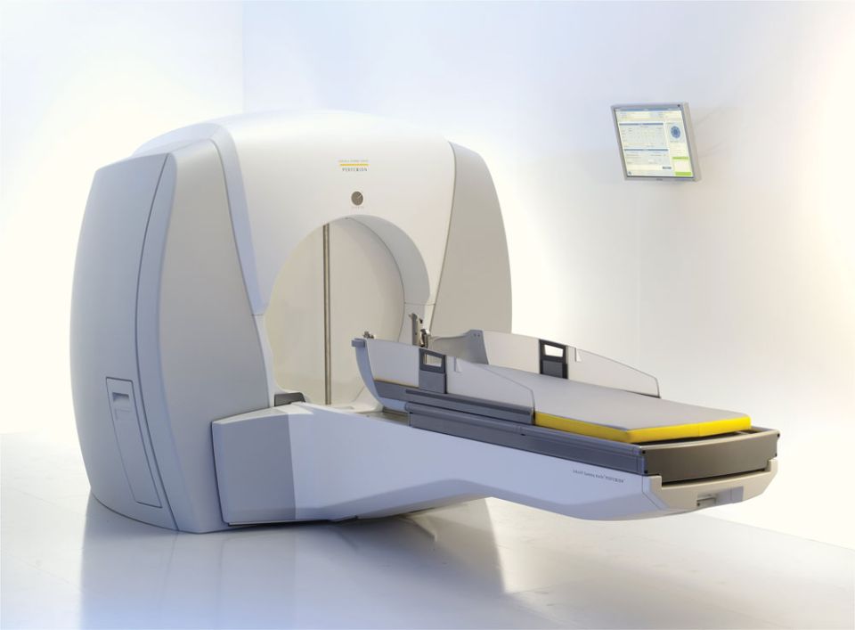
Gamma Knife is a radiosurgery system that focuses gamma rays from cobalt-60 sources onto a target inside the skull, delivering an effective dose in a single session while protecting the surrounding normal tissue.
Gamma Knife is a radiosurgery system that uses stereotactic methods to focus gamma rays emitted from cobalt-60 sources onto a defined target, delivering an effective dose in a single session while exposing the surrounding normal tissue to as little radiation as possible. It can be used only for lesions located inside the skull.
Its key difference from radiotherapy is the ability to deliver a high dose in a single session while protecting normal tissue.
Gamma Knife radiosurgery has removed the need for open brain surgery in many conditions and has provided a safe and effective treatment option for many patients.
History
Radiosurgery was first described in 1951 by the neurosurgeon Lars Leksell at the Karolinska Institute, and the first treatment was carried out in 1968. With advances in technology and software it has become increasingly widespread, and as the level of evidence has grown it has become a preferred treatment method worldwide.
Neuroradiosurgery in Turkey dates back roughly twenty years. The country's second centre was founded in 2004 in Ankara, at the Department of Neurosurgery of Gazi University Faculty of Medicine, by Prof. Dr. Aydın Paşaoğlu, initially with a Gamma Knife Model C. A Gamma Knife Perfexion was installed in 2014, and the centre continues its work with more advanced technology. By 2020 more than 9,000 patients had been treated there.
The safety and effectiveness of Gamma Knife for certain brain conditions has been demonstrated worldwide through experience with more than one million patients and more than 15,000 publications. It has replaced surgery for many conditions and is accepted as an adjunct to surgery in many others.
Gamma Knife systems have evolved considerably with advancing technology, but the basic operating principle and philosophy have remained the same.
Gamma Knife can deliver target-specific three-dimensional treatment with an accuracy of 0.1 mm. Treatment time varies with the dose and the volume of the target.
Patients are observed for a few hours after treatment and then discharged. Apart from fitting the frame under local anaesthesia and placing an intravenous line, the procedure requires no surgical intervention or special medication. After treatment, patients are followed with periodic MRI scans.
The Gamma Knife system
Gamma Knife consists of five separate units:
- Radiation unit: Different models contain different numbers of cobalt sources. In models other than Perfexion and Icon, 201 collimators and their corresponding cobalt sources are arranged in a hemisphere so that they target its centre point.
- Collimator system: The collimators set the volume of tissue exposed to each beam, allowing radiation fields to be shaped to the lesion. In Perfexion and Icon the collimators are arranged on sector rails.
- Patient positioning system: The couch on which the patient lies, which moves the patient automatically into the radiation unit.
- Stereotactic frame: A Leksell stereotactic frame is fixed to the head under local anaesthesia. It acts as a reference ruler, allowing the coordinates (X, Y and Z) of the target point to be determined numerically. It is fitted before imaging and stays in place until the end of treatment; marker boxes for CT, MRI and DSA are attached to it.
- Treatment planning system: A separate computer system that imports the patient's CT, MRI and DSA images, defines the target and the dose, and transfers the treatment plan to the treatment unit.
Stages of Gamma Knife treatment
Gamma Knife treatment can usually be completed as an outpatient procedure in about 2–3 hours, without hospital admission. Starting with frame placement and ending with dose delivery, it involves the following steps:
- Placement of the stereotactic frame
- Neuroradiological imaging: CT, MRI and/or DSA are obtained with the frame in place.
- Dose planning: The stereotactic images are loaded into the planning computer, the target is defined and the dose is shaped to fit the target. The doses received by nearby critical structures are measured and the plan is adjusted to increase safety. The system includes numerous automatic checks designed to prevent operator or system errors.
- Dose delivery: Treatment is delivered according to the plan transferred to the treatment unit. No pain is felt while the radiation is being delivered.
Which conditions can be treated with Gamma Knife radiosurgery?
Most benign and malignant intracranial tumors that do not cause significant compression of the nervous system (roughly smaller than 3–4 cm), including:
- Meningioma
- Vestibular / trigeminal schwannoma
- Pituitary adenoma
- Craniopharyngioma
- Metastases
- Glial tumors
- Hemangioblastoma
- Chordoma
- Glomus jugulare tumors
Cerebrovascular diseases:
- Arteriovenous malformation
- Cavernoma
- Cavernous sinus hemangioma
Other:
- Trigeminal neuralgia
- Parkinson's disease and tremor
- Obsessive–compulsive disorder
